Ankle Dorsiflexion: What Restricts It, How to Test It, and How to Rebuild It
By Brian Murray
Every coach who’s spent real time under the bar with people eventually runs into the same wall. Someone can’t get their squat below parallel no matter how much hip mobility work they do, and the fix isn’t in the hip at all. It’s six inches lower, at a joint nobody thinks about until it stops cooperating.
Ankle dorsiflexion is the motion of the shin traveling forward over a planted foot. It’s what has to happen every time you squat, lunge, walk downhill, or absorb a landing. It’s also probably the most underdiagnosed restriction in general population training, because it hides behind other joints. People blame their hips for a shallow squat. They blame their quads for knee pain. Rarely does anyone check the ankle first, even though the ankle is often the actual ceiling.
What Dorsiflexion Actually Is, and Why the Test You Use Matters
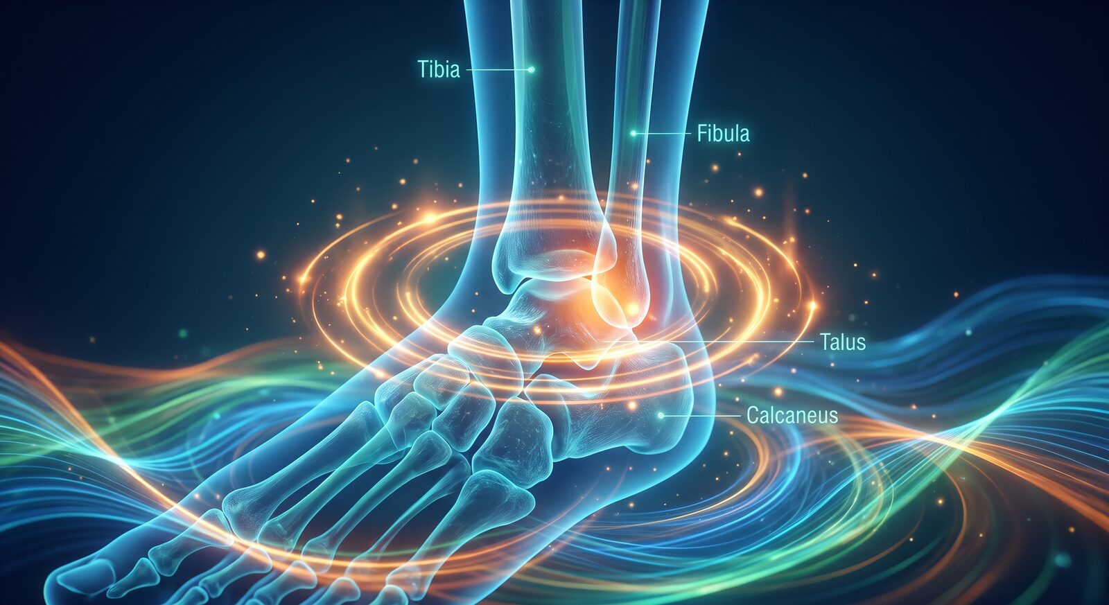
The talocrural joint is formed by the talus sitting inside the mortise created by the tibia and fibula. The talar dome isn’t a simple cylinder. It’s wedge-shaped, wider at the front than the back, which means full dorsiflexion requires the mortise itself to widen slightly to let that wider section through (8). That widening is small, roughly a millimeter, produced by a couple degrees of external rotation at the fibula (9). Most people never think about the fibula as part of an ankle mobility problem. It usually is one, in a small but real way.
There are also two ways to measure the range itself, and they don’t measure the same thing. Non-weight-bearing dorsiflexion, done lying down with someone else moving your foot, tells you about passive tissue length. Weight-bearing dorsiflexion, tested with the knee driving forward over a planted foot (the weight-bearing lunge test, or WBLT), tells you what’s actually available under load. Research comparing the two found the weight-bearing measurement predicted compensation patterns during squatting, while the non-weight-bearing test didn’t (1). If you only ever test dorsiflexion on a table, you’re measuring the wrong thing for a training context.
Normal WBLT values sit above roughly 44 degrees in a lot of the literature, with values under 30 degrees flagged as a meaningful functional deficit (4). That’s a wide window, and individual variation inside it is real. But someone testing well under 30 isn’t expressing a preference. That’s a restriction with downstream consequences.
Where the Restriction Actually Comes From
This is the part most internet advice skips straight past on its way to prescribing calf stretches. Dorsiflexion can be limited by at least four different structures, and they don’t respond to the same fix.
The first is the joint capsule and the posterior glide of the talus. Dorsiflexion requires the talus to roll and glide backward inside the mortise as the shin moves forward. If that glide is restricted, calf length is irrelevant, because the limiter isn’t muscular at all. This is the structure manual therapy research keeps showing improvement in. Posterior talar glide mobilizations have produced measurable, sometimes fairly durable gains in dorsiflexion in people with a history of ankle injury (7).
The second is that small but real mortise mobility described above. A stiff syndesmosis, often from an old high ankle sprain or just general stiffness through the lower leg, can cap the last few degrees of range even when the capsule and calf are both cooperating. It’s rarely the whole story on its own, but it’s part of why some ankles stay stubborn after everything else has been addressed.
The third and fourth are the two calf muscles, and they restrict dorsiflexion through different mechanisms. The gastrocnemius crosses both the knee and the ankle, so it goes slack when the knee bends. The soleus only crosses the ankle, so knee position doesn’t change its tension. This is the entire logic behind the Silfverskiold test, which compares dorsiflexion with the knee straight against dorsiflexion with the knee bent to 90 degrees. If range improves meaningfully with the knee bent, the gastrocnemius is the limiting structure. If the number barely moves, the restriction sits in the soleus, the capsule, or the mortise instead. The test has held up well diagnostically, with sensitivity and specificity both in the high 80s and 90s against goniometric measurement in the podiatric literature (11). It’s also worth coaching this distinction because the injury associations differ. Gastrocnemius-dominant restriction tracks with ankle sprain history, stress fractures, and shin splints. Soleus-dominant restriction tracks more with plantar fasciitis and Achilles tendinopathy (10). Different pattern, different downstream risk, same test to sort it out.
Then there’s the structure that gets missed most often, because it isn’t soft tissue at all. Anterior ankle impingement is a bony block, osteophyte formation at the front of the tibia and talus, usually built up from repetitive terminal dorsiflexion loading or a history of ankle sprains (13, 14). It shows up constantly in soccer players, dancers, and jumpers for exactly that reason. This one has a distinct feel on assessment. Instead of a springy, stretch-like resistance at end range, there’s a hard, sometimes sharp stop, often with localized pain right at the front of the joint. No amount of stretching or mobilizing changes bone. If that’s what you’re feeling under your hands, the right move is a referral, not a longer hold.
Why This Shows Up in Your Squat Before It Shows Up Anywhere Else
Walking doesn’t ask much of the ankle. Normal gait requires somewhere around 5 to 15 degrees of dorsiflexion during stance, with some research putting the functional minimum around 10 degrees (12). That’s exactly why a real restriction can go unnoticed for years. Someone can walk around fine every day and still be sitting well under the range they need the moment they load a squat, take stairs two at a time, or run uphill, since jogging demands noticeably more range than walking does (12).
The squat is where it gets exposed fastest, because it’s the highest closed-chain demand most people put on the joint. A parallel back squat requires somewhere around 23 to 31 degrees of dorsiflexion depending on setup and footwear (3). People who test below that on the WBLT don’t just lose depth. They change the whole strategy of the lift, shifting load into greater hip and lumbar flexion and a more pronounced forward trunk lean to find the range somewhere else in the chain (2). One study found that roughly every extra 6.5 degrees of weight-bearing dorsiflexion translated to about 10 percent more squat depth (3). That’s not a small relationship.
It doesn’t stop at depth. Limited dorsiflexion has also been linked to altered knee mechanics during squatting and single-leg tasks, including greater knee valgus displacement, which matters for anyone tracking ACL injury risk factors (1). And reduced dorsiflexion has been associated with a higher rate of Achilles tendinopathy, with one prospective study putting the risk at roughly two and a half times higher in people with the least available range (4). None of this means restricted dorsiflexion causes those outcomes on its own. Bodies are more complicated than a single-joint explanation. But the pattern is consistent enough that checking the ankle before rebuilding a squat, a landing mechanic, or a running gait isn’t optional. It’s the first place to look.
Where the Evidence Gets Messy
I want to be straight about the limits here, because this is exactly the kind of topic that gets oversold. Joint mobilization research on dorsiflexion is promising but not settled. A systematic review with meta-analysis on ankle mobilization found a statistically significant pooled improvement in dorsiflexion, but that significance disappeared once the analysis was restricted to only the higher quality trials (5). That’s not a reason to drop the technique. It’s a reason to say the effect is real in some people and contexts, and probably smaller and less reliable than the confident version of this claim you’ll see elsewhere.
Static calf stretching has a similar story. A systematic review found static stretching does increase dorsiflexion, with effects in the two to three degree range depending on duration, and the researchers considered the results at 15 to 30 minutes of stretching robust (6). Two to three degrees is real, but it’s small next to a 44 degree normal range, and nowhere close to what you’d need if the actual limiter is capsular, bony, or sitting in the mortise instead of the muscle. This is the same lesson from the isometrics piece showing up from a different angle. The dose that produces change is usually longer and more specific than what people are actually doing, and the method has to match the structure you’re trying to change, not just the joint.
How to Actually Test and Rebuild It
Start with the weight-bearing lunge test against a wall. Foot a few inches back, knee driving forward to touch the wall without the heel lifting, and walk the foot back until you find the true limit. That’s the only version of this test worth trusting for training decisions, since it’s the one that actually predicts what happens under load (1).
Pay attention to what the end range feels like, not just the number. A hard, sudden stop with localized pain at the front of the ankle is worth referring out before you program anything into it. That’s the impingement pattern, and loading into it repeatedly is how it gets worse, not better.
If the end range feels soft and stretch-like instead, run the Silfverskiold comparison. Straight knee, then bent knee, same wall test both times. A meaningful improvement with the knee bent points to the gastrocnemius, and the fix is loaded gastrocnemius-length work, a straight-leg calf stretch held under real tension rather than the twenty second version most people default to. Little to no change between the two positions points away from the muscle and toward the capsule or the mortise, which is your cue to work joint mobilization instead, whether that’s a banded ankle distraction into dorsiflexion or hands-on work if there’s an old sprain in the history.
CARs at the ankle still have a place here, the same way they do everywhere else on this site. They’re not how you build the range. They’re how you check whether the range you’ve built is still there and still under control (see What CARs Are Actually For). If you want to see this whole sequence run start to finish, from CARs into PAILs and RAILs into loaded reinforcement, we put out a free ankle mobility foundations class on YouTube that walks through exactly this progression at the ankle, no gear required and no cost to watch it. Once dorsiflexion improves on the wall test, the last step is putting it back into the pattern it was missing from. Loaded ankle dorsiflexion drills, deficit reverse lunges, and knee-forward isometric holds at end range all put the new range under the kind of demand it needs to survive contact with an actual lift.
Restricted dorsiflexion is rarely the whole story behind a shallow squat or a cranky knee. But it’s cheap to test, specific enough to actually fix once you know which of the four structures is limiting it, and it’s the joint most people skip past on their way to blaming somewhere else. Check it first. You might save yourself a lot of hip mobility work that was never going to solve the actual problem.
References
- Dill KE, Begalle RL, Frank BS, Zinder SM, Padua DA. Altered knee and ankle kinematics during squatting in those with limited weight-bearing-lunge ankle-dorsiflexion range of motion. J Athl Train. 2014.
- Effect of limited ankle dorsiflexion on lower limbs and trunk kinematics during squatting. ScienceDirect, 2025.
- Role of ankle dorsiflexion in sports performance and injury risk: a narrative review. EJGM.
- Comprehensive corrective exercise program improves ankle function in female athletes with limited weight-bearing ankle dorsiflexion. PMC, 2024.
- Effect of joint mobilization on chronic instability of the ankle: a systematic review with meta-analysis. Arch Med Deporte, 2022.
- Does stretching increase ankle dorsiflexion range of motion? A systematic review.
- Posterior talar mobilizations improve ankle pain, dorsiflexion and functional capacity (review of Silva et al. 2017). Brookbush Institute.
- Talocrural and subtalar joint anatomy and syndesmosis mechanics. PMC.
- Fibular rotation and mortise widening during dorsiflexion. OrthoRacle.
- Isolated gastrocnemius contracture: diagnostic criteria and Silfverskiold test performance. Foot & Ankle International.
- Examination techniques of foot and ankle special tests, including the Silfverskiold test. World J Orthop, 2017.
- Ankle dorsiflexion requirements during walking and jogging gait. Frontiers in Neurology, 2023.
- Anterior Ankle Impingement Syndrome overview.
- Anterior bony impingement of the ankle: etiology and imaging. PMC.